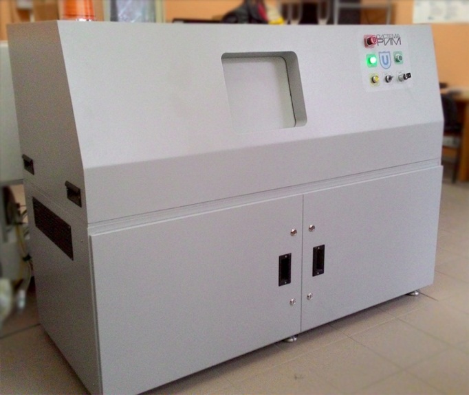
DIGITAL X-RAY MICROTOMOGRAPH
Дата 01.12.2017 16:40:00 | Раздел: The Center for Excellence "Intellectual Technical Systems"
| Digital X-Ray Microtomograph is used to study the spatial structure of the materials, crystals, organic and inorganic objects with dimensions up 20 x 20 x 20 mm and a resolution of 1 to 13 microns
Specifications
- Distinguishability: 1-13 microns
- X-ray source: smoothly adjustable from 20 to 160 kV, anode current: 0 – 250 mkA, 10 Watt, focal spot size of <5 microns (≅ 4 watts), air-cooled
- X-ray detector: 4872 x 3248, size of single cell 7,4 x 7,4 microns
- The recovery time of three-dimensional images (1 cm^3): 10 m
- Analysis of three-dimensional images (1 cm^3): 30 m
Benefits
- High-precision positioning system that can ensure the positioning of the object with an accuracy of ± 1 mkm
- Full automation of the X-ray microtomograph that does not require any user intervention in the process of building a 3D-model of the object
- Built-in algorithms for the analysis and classification of the internal structure and the defects of the object
- Built-in algorithms for pre-processing of digital data and undistorted compression in order to save computing resources
The object of intellectual property: 250580, 2476825
Delivery time: 3-6 months
The price is negotiable
|
|